Quanterix Akoya PhenoImager HT 2.0
Quanterix Akoya PhenoImager HT 2.0
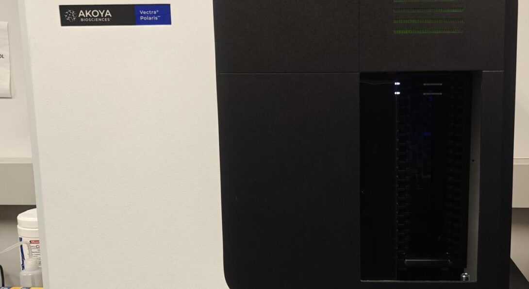
The Quanterix Akoya PhenoImager HT 2 is a whole-slide imaging system built for quantitative spatial phenotyping and high-throughput tissue analysis.
Core System Specifications
- Optical Magnification: Supports digital whole-slide scanning at 10x (1.0 μm/pixel), 20x (0.5 μm/pixel), and 40x (0.25 μm/pixel) resolutions.
- Slide Capacity and Automation: Features a touchless, automated loader that accommodates up to 80 slides simultaneously. The system utilizes continuous loading technology to permit slide replacement during active scanning operations.
- Imaging Modalities (Brightfield, MSI or Motif whole slide scans): Acquires images in either brightfield or fluorescence formats from tissue sections or tissue microarrays (TMAs). For fluorescence, the system uses either long-pass filters in conjunction with a liquid crystal tunable filter (LCTF) or a set of narrow band filters to collect multispectral images.
- Software Integration: Links to inForm and phenoptr semi-automated tissue analysis packages, which use machine-learning algorithms to execute tissue segmentation, cell segmentation, and phenotypic scoring. Whole slide unmixed multispectral images can be created and analyzed with Indica Labs Halo.
- File Output: Exports data in the standardized Akoya whole-slide multi-resolution tiled format (.QPTIFF) or individual multispectral field formats (.im3).
7 Color Multispectral Imaging and Onboard Unmixing
The platform integrates reference library spectral profiles directly into the scanning sequence:
- Protocol Loading: The generated Spectral Library is uploaded into the instrument software prior to beginning a scanning batch.
- Multispectral Acquisition: The LCTF system records raw multiplexed emission data across multiple discrete wavelength bands during the tissue scan.
- Linear Matrix Solving: The system software applies a linear unmixing algorithm on a pixel-by-pixel basis. The total measured light intensity at each pixel is calculated as a linear combination of the individual dye components from the library.
- Real-Time Processing: The instrument processes these matrix equations dynamically alongside real-time image stitching as data capture proceeds across the slide.
9 Color Multispectral Imaging Capabilities
The PhenoImager HT integrates a hardware-based Liquid Crystal Tunable Filter (LCTF) in its emission optical path to manage spectral overlap in multiplexed panels.
- Wavelength Spectrum: Operates across a multispectral range of 440 nm to 780 nm, capturing emission data across more than 40 discrete wavelength bands.
- Multiplex Capacity: Isolates up to 9 distinct colors (typically DAPI plus 6 to 8 fluorescent biomarkers, such as Opal dye sets) within a single tissue section.
- Spectral Unmixing: Leverages established reference libraries (derived from single-stained and unstained tissues) to calculate and separate overlapping emission signatures. The instrument software processes this math on a pixel-by-pixel basis.
- Autofluorescence Isolation: Treats tissue-specific background autofluorescence as an independent channel during the unmixing calculations. This separates the background noise from target fluorophore signals to increase the signal-to-noise ratio.
Compatible Dyes
| Scan Band | Opal Dye | Compatible Alexa Dye | Compatible Cyanine Dye | Compatible Other Dye |
|---|---|---|---|---|
| DAPI | Spectral DAPI | DAPI | ||
| Opal 480 | Opal 480 | AF405(!) | Coumarin 6 | |
| Opal 520 | Opal 520 | AF488 | Cy2 | FITC |
| Opal 570 | Opal 570 | AF555 | Cy3 | TRITC |
| Opal 620 | Opal 620 | AF594 | Cy3.5 | Texas Red |
| Opal 690 | Opal 690 | AF660, AF680(!) | Cy5.5 | |
| Opal 780 | Opal 780 | AF750 | Cy7 |